Department Microstructure and Residual Stress Analysis
Imaging & diffraction setup
The white-beam (6 - 120 keV) of up to 4 x 4 mm2 section and high photon flux available at EDDI is well suited for time-resolved in-situ experiments. Radiograms with up to 1 kfps, tomograms with 2 µm spatial resolution and whole diffractograms can be obtained in 1 s simultaneously. This combined with sample environments like heating stages or mechanical testing machines either brought by users or readily available at the beamline open new possibilities for in-situ studies.
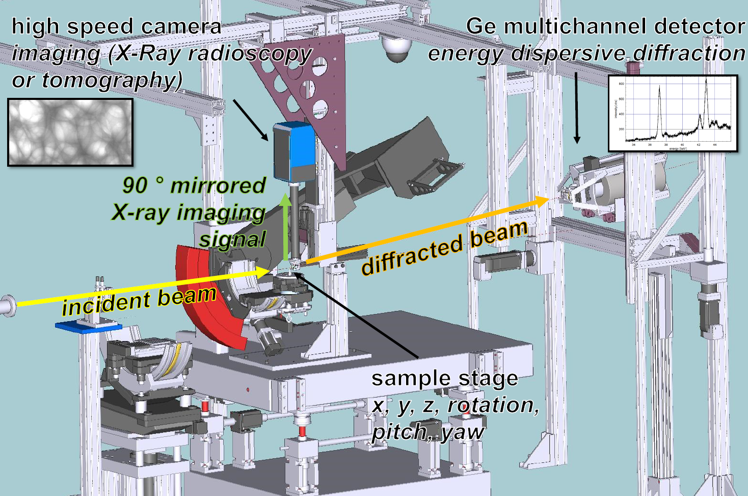
The diffracted beam detected at 2θ = 6° by a Ge-Multichannel analyser (Canberra GL0110). The direct transmitted beam is converted into visible light (LuAG scintillator, 200 µm), which is reflected by a mirror and recorded by a fast PCO Dimax CMOS camera (2016 x 2016 pixels and 11 x 11 µm2 pixel size, > 7 kfps acquisition by 1000 x 1000 pixel).).
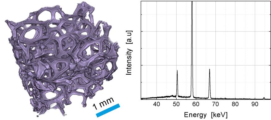
Tomogram and diffractogram of a Ni-based open-cell foam both acquired simultaneously in 10 s.
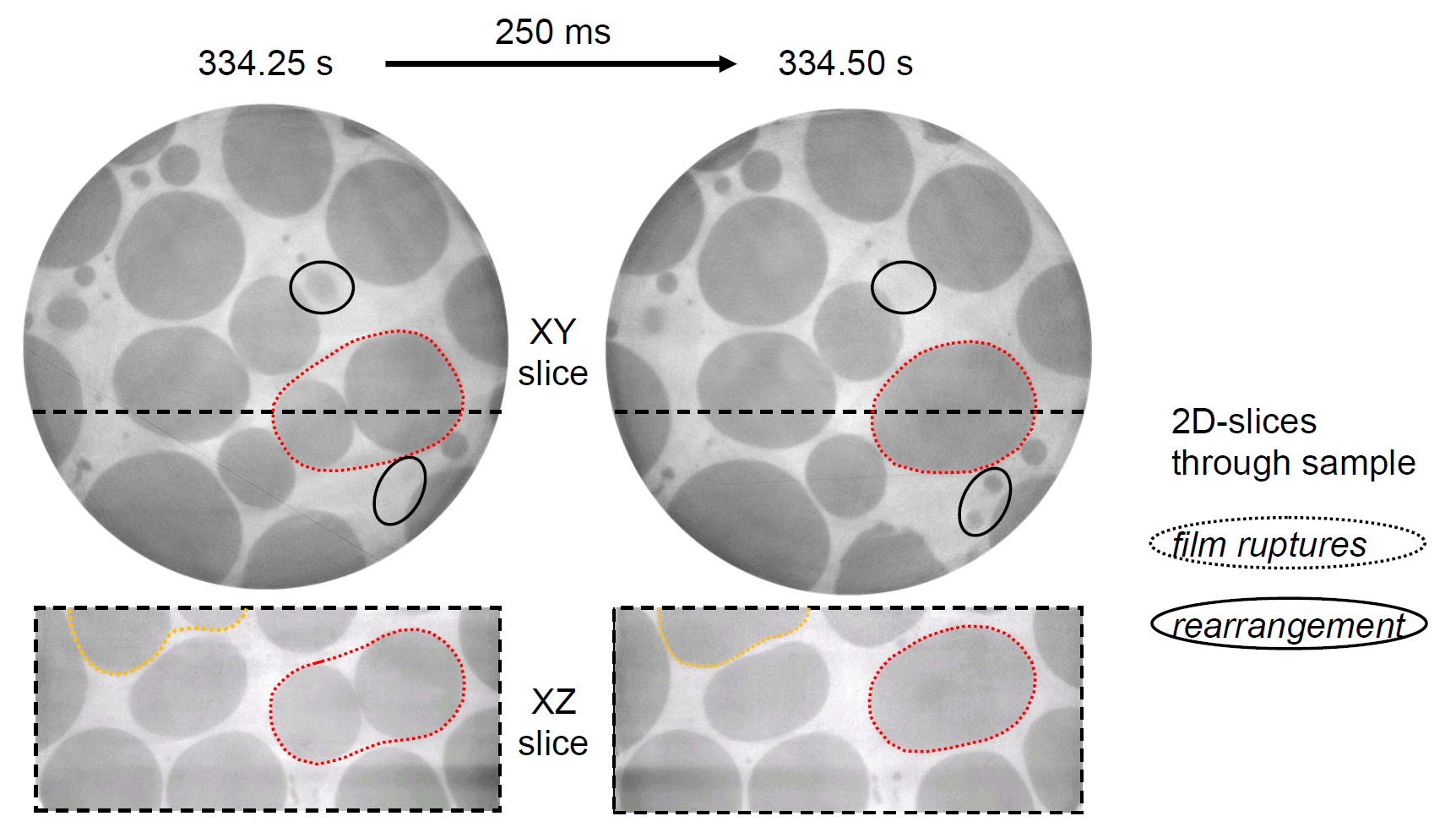
Slices of a liquid metal foam extracted from a tomogram series acquired in 0.25 s (4 tomos/s) showing the structure evolution.