2.1. Quantum materials for energy
2.1.4. Nanoscale materials
At the nanoscale, materials exhibit novel physical, chemical, electrical, magnetic, and optical properties that can be exploited for a wide variety of applications ranging from catalysis to magnetic data storage. An understanding of static and dynamic properties of such low-dimensional structures is only accessible to spectro-microscopic tools capable of appropriate lateral and temporal resolution. Synchrotron based imaging techniques, such as coherent x-ray scattering (CXRS) or photoemission electron microscopy (PEEM), offer a unique toolbox for nanoscale science. The combination of multi-specific contrast with temporal and lateral resolution at the relevant time and length scales enables these microprobes to follow chemical reactions in a catalyst or image the magnetic switching in a nanoparticle. Complemented by structural analysis, such as transmission electron microscopy (TEM), they provide unmatched insight into the structure-function relationship at the nanometer level.
It has been demonstrated that the electronic structure and magnetic response of the same single ferromagnetic nanoparticle can be correlated with the morphology and crystal structure using x-ray photoemission microscopy in combination with electron microscopy (Figure 4). Magnetic states and interactions can be analyzed for different particle arrangements. The element-specific electronic structure can be probed and correlated with the changes of magnetic properties. This approach opens new possibilities for a deeper understanding of the collective response of magnetic nanohybrids in multifunctional materials and in nanomagnetic colloidal suspensions used in biomedical and engineering technologies.
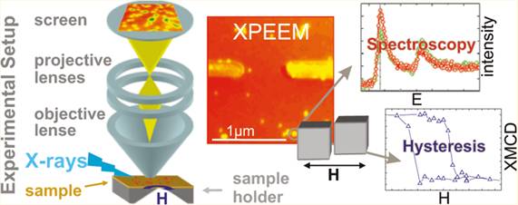
Figure 4. Magnetic imaging and spectromicroscopy of individual Fenanocubes in an applied magnetic field of up to 33 mT. (Courtesy F. Kronast, HZB).
Another important application is on materials that are inhomogeneous on the nanoscale due to spontaneously developing order phenomena or coexistence of different phases. Both kind of effects have clear influence on macroscopic properties and affect hence functionality. Techniques that have both temporal and spatial resolution can unravel on a nanoscale how functionality can best be achieved. In addition to the aforementioned imaging technique, x-ray photon correlation spectroscopy will be an essential tool.
BESSY-VSR offers X-ray pulses at variable pulse length in combination with variable filling patterns with, e.g., alternating patterns of ultra-short and long pulses. The availability of ultra-short X-ray pulses with a pulse duration below one picosecond will provide experimental access to new phenomena such as direct spin-excitations in individual nanoparticles or magnetic fluctuations in particle ensembles.
The variability of BESSY-VSR could also be used to solve a problem of electron-detection-based imaging such as PEEM at current X-ray sources. Above a certain X-ray intensity their resolution is limited by space charging effects. Recent studies at BESSY II and Elettra (Trieste, Italy) demonstrate that even with the present X-ray pulse duration and intensity, the lateral resolution in PEEM can be limited by space charging. With BESSY-VSR we could overcome this space charging with particularly long X-ray pulses. If technically feasible, this would pave the way towards nm resolution X-PEEM, as predicted by electron optical simulations.
Shorter X-ray pulses, in turn, as provided by BESSY-VSR would enable new insights with ultrafast hard X-ray diffraction experiments:
Nonlinear phononics: The dynamics of phonons in an anharmonic crystal potential
The decomposition of atomic motion in phonon modes is a fundamental concept in solid state physics. Phonons describe elementary processes and excitations, such as heat transport, sound propagation, pyro- and pizo-electric processes, phase transitions and others. For structural phase transitions with unit cell doubling the zone boundary phonons are relevant, which have a decay constant in the 1 picosecond range. Coherent phonons can be generated and probed by ultrashort light pulses. The accessible phonon wavevector q in the probe process is however very limited for optical wavelengths. It is therefore advantageous to reduce the probe wavelength by using radiation in the hard X-ray regime.
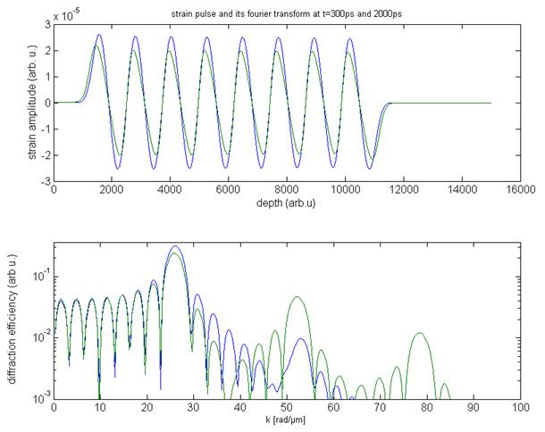
Figure 5. (upper panel) Nonlinear phononics: the initial acoustic strain pattern (blue) is distorted (green) after propagation in an anharmonic crystal. (lower panel) The excitation of new phonon modes can be observed in reciprocal space by ultrafast X-ray diffraction. (Courtesy P. Gaal, R. Shayduk, M. Bargheer, Universität Potsdam).
In particular, for the study of anharmonic effects, e.g. the decay of coherent sound waves due to phonon-phonon interactions, the sensitivity to a large set of phonon wavevectors and a sufficient temporal resolution is required. The decay time constant for acoustic phonons with a frequency of 140 GHz is approximately 100 picoseconds [1]. In general, the phonon decay follows a 1/w2-rule, i.e. high-frequency phonons and localized lattice distortions decay on a sub - 100 picosecond timescale [2]. Such high-frequency modes can be probed by Ultrafast X-Ray Diffraction (UXRD) techniques, due to the larger k-vector of X-rays compared to optical light (Figure 5) [3]. However, UXRD experiments on the decay of phonons are rare, since they rely on ultrastable and brilliant X-ray probe pulses that can be delivered uniquely by accelerator-based sources, which typically deliver a temporal resolution of only 100 picoseconds [4].
The generation of large strain amplitudes leads to nonlinear propagation effects and to the excitation of new phonon modes due to anharmonic phonon interaction [5]. A better understanding of nonlinear phononics could pave the way to new devices, e.g. modulators and switches [6,7].
For such studies, an increase in temporal resolution as provided by BESSYVSR for accelerator-based UXRD experiments is essential.
References for section 2.1.4.
[1] M. Herzog et al., APL 100, 09401 (2012).
[2] A. Akhiezer, J. Phys. (USSR) 1, 277 (1939).
[3] R. Shayduk et al. ,(submitted).
[4] H. Navirian et al., Rev. Sci. Instr. 83, 063303 (2012).
[5] A. Bojahr et al., Phys. Rev. B 86, 144306 (2012).
[6] P. Gaal et al., APL 101, 243106 (2012).
[7] P. Gaal et al., (submitted).