On-site Trainings
(30 March - 2 April, 2026)
This is a list of the synchrotron–based and laboratory-based experiments that are offered as on-site trainings. Please choose up to two experiments of your preference.
Training 01
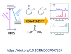
TD-DFT simulations of K-edge resonant inelastic X-ray scattering
Vinícius Vaz da Cruz, Wagner Ribeiro da Silva Neto
In this exercise, you will be exposed to the basic concepts of molecular modelling and ab initio interpretation of experimental data. The tutorial ranges from molecular geometry optimizations, orbital visualization to spectral calculations, focusing on the resonant inelastic X-ray scattering process (RIXS). The tasks will focus on applying simulations of molecular electronic structure using Density Functional Theory (DFT) and its time dependent extension (TD-DFT) to access excited electronic states. We will investigate both occupied and unoccupied molecular orbitals that define bonding in molecular systems and discuss how they are probed within RIXS and how modelling allows drawing deeper insight from the measured spectra. The simulations will be carried out using the Orca quantum chemistry package for electronic structure calculations and Avogadro and Molden will be used for visualization of the results.
Training 02
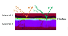
Accessing deeply buried interfaces via Hard X-ray Photoelectron Spectroscopy
Roberto Félix Duarte, Marcus Bär
The complexity of energy conversion devices, which often comprise a multitude of layers, interfaces, surfaces, elements, impurities, etc., dictates that it is of crucial importance to characterize and understand the chemical and electronic structure of each component both individually and as part of the larger system. In this module, students will get an introduction to hard x-ray photoelectron spectroscopy (HAXPES), an extraordinarily powerful method to determine the chemical and electronic characteristics of buried interfaces. By employing a range of excitation energies, the probing depth of the HAXPES measurements can be tuned to focus on the very surface of a sample or deeper into its near-surface region. This approach will serve as a straightforward way to carry out elemental depth profile analyses of an initially (i.e., at room temperature) inert heterointerface (i.e., “Material 1”/“Material 2”), an efficient alternative to measuring a sample series with different “Material 1” layer thicknesses. Furthermore, the reactivity of the investigated heterointerface as a function of sample temperature will be assessed by monitoring various element intermixing during in situ annealing treatment. The impact of thermally induced diffusion mechanisms on the electronic interface structure will also be explored. This proposed course of action provides a more comprehensive picture than characterizing the chemical and electronic structure of sample series treated ex situ by different annealing temperatures. The experiments will be conducted at the High Kinetic Energy Photoelectron Spectrometer (HiKE) endstation located at the KMC-1 beamline of the BESSY II light source.
Training 03
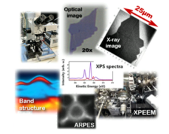
2D materials under the X-ray microscope
Alevtina Smekhova, Florian Kronast
Atomically thin two-dimensional (2D) materials demonstrate intriguing physical, chemical, optical, electronic and magnetic properties, and are currently considered as promising candidates for the key blocks in the next-generation quantum and optoelectronic devices. Nowadays, many 2D materials can be organized in various vertically stacked van der Waals (vdW) heterostructures with the macroscopic lateral dimensions and stabilized against environmental degradation by different capping layers. In the future, these materials would allow substantial miniaturization of modern electronic devices along with significant expansion in their functionalities. In our training we invite participants to prepare 2D materials by mechanical exfoliation of vdW crystals in air with a possible assembly of a vdW heterostructure. The prepared 2D samples will be first evaluated by optical and eventually atomic-force microscopy, and then room-temperature electronic properties of particular configurations will be investigated by X-ray Photoemission Electron Microscopy (XPEEM) using element-specific X-ray techniques like XAS, XPS, and micro-ARPES. The stability of the prepared 2D samples and changes at their surfaces under heating can be checked in addition.
Training 04
Multiple Energy Anomalous X-ray Diffraction (MEAD) on quaternary semiconductors
Daniel Többens
In this experiment, for example Kesterite‐type Cu2ZnSnS4 and Stannite‐type Cu2FeSnS4 will be investigated by Multiple Energy Anomalous X‐ray Diffraction (MEAD). For these spectra over the X‐ray absorption edges of Cu (8979 eV) and Zn (9659 eV) will be acquired at the KMC-2 Diffraction station (or MySpot beamline). The energy dependence of intensity corresponding to Bragg peaks 011 and 110 shows very distinctive differences depending on the distribution of copper and zinc. This allows clear and immediate distinction between kesterite‐type (real) and stannite‐type (once speculated, but disproved) crystal structure. Distinction between ordered and disordered Cu2ZnSnS4 is less obvious and ambiguous in this experiment, demonstrating also the limitations of this technique.
Training 05
Monitoring buried interface formation using in-system laboratory- and synchrotron-based photoelectron spectroscopy
Johannes Frisch, Regan Wilks, Marcus Bär
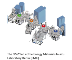
The interfaces formed in multilayer thin films are the key to their functionality, and their characterisation in non-destructive manners is an area of great interest. The SISSY lab at the Energy Materials In-situ Laboratory Berlin (EMIL) features an advanced surface-sensitive characterization system which is connected via ultra-high vacuum transfer to deposition chambers. Avoiding exposure to ambient conditions helps guarantee that deposition of a film can be interrupted and restarted at various times with only a small influence on the final film composition. This experiment will involve stepwise, in-vacuum deposition of thin films combined with a series of surface sensitive x-ray (XPS) and ultraviolet (UPS) photoelectron spectroscopy measurements. XPS measurements will describe film thickness, layer closure, chemical structure, and shifts in energy levels as the interface is formed. UPS measurements of the valence band maximum position and work function will complete the study of the electronic levels. The set of measurements will allow a complete picture of the energy level alignment at the interface to be built up and used to understand the behaviour of the resulting device.
Training 06

XANES in Standing Wave Geometry to study a W/Si layered interface
Ivo Zizak
An interface between the W and Si Layer will be studied at the MySpot beamline. The W layer is embedded into a waveguide between Au and Mo layers, with a Si spacer. The exact structure is shown in the figure. XANES spectra will be collected for different distributions of the electric field, demonstrating the depth sensitivity. From the 2.2 nm thick W layer, comparison is done between the spectra measured in the middle of the layer (metallic W) and at the interface to Si. In this experiment, we will first measure the reflectometry and fluorescence from the sample. Evaluation of these spectra will help to estimate the distribution of the X‐ray in depth of the sample. Results will be compared to a simulation and differences will be discussed. Two geometries are selected: i) X‐ray intensity is concentrated in the middle of the layer, giving pure metallic W spectrum, and ii) maximal intensity is at the interface between W and Si, giving spectrum specific for WSi alloy. According to the results of the first part, we will be able to set the parameters for an XANES experiment. Dependency between the electric field pattern and wavelength, as well as the necessary angle correction will be discussed. The data will be loaded and viewed using Athena software suite. Provided literature will be used to be compared with the results
Training 07
Resonant Inelastic X-ray Scattering (RIXS)
Maximilian Kusch, Katarzyna Siewierska, Marco Kliem
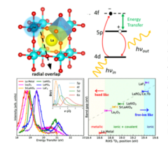
Low energy excitations in functional materials are important to reveal their intrinsic properties. In this experiment, we are going to use photon-in-photon-out technique at EUV and soft-x-ray regions (30-270 eV range) to unveil the bandgap in lanthanum-containing insulators [1]. The participants will learn from sample preparation, code-based measuring techniques to data analysis. The solid samples will be probed by X-ray absorption spectroscopy (XAS) and resonant inelastic x-ray scattering (RIXS), which will be conducted at the dedicated meV-RIXS experimental station [2]. The high-resolution setup serves as a powerful tool to unambiguously determine the electronic transition and its coupled degree of freedom [3]. With the precise determination of the peak position, we will use the atomic 5p-4f transition as a local atomic sensor. Based on the chemical shift observed, we will see how this valence transition is perturbed by the band(gap) formation and determine the bandgap of the unknown samples by the established linear regression.
[1] C.-Y. Liu et al., J. Phys. Chem. C 2023, 127, 23, 11111-11118. https://doi.org/10.1021/acs.jpcc.3c02011
Training 08
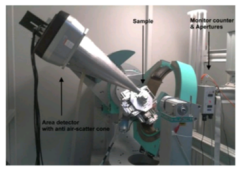
MX beamlines
Manfred S. Weiss, Uwe Mueller, Frank Lennartz, Tatjana Barthel
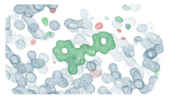
We will prepare crystals of the sweet protein thaumatin beforehand. During the practical training, the students will prepare the crystals for the diffraction experiment, by mounting them in a nylon loop and immersing them in Liquid N2. A diffraction experiment will be carried out at one of the MX beamlines. The data will be processed and the structure determined by molecular replacement. A small molecule binding to the surface of thaumatin will be identified by difference density analysis.
Training 09
3D/4D hard X-ray Imaging of Batteries
Ralf F. Ziesche, Henning Markötter
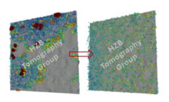
In recent years, Li-ion batteries and their potential successors have become one of the most important energy storage devices for portable technology such as mobile phones and electric cars. However, the real available gravimetric/volumetric energy density and rate performance are still far from the theoretical ones. Multi-dimensional characterisation techniques are the key to a deep understanding of battery operation and performance. Hard X-ray imaging provides valuable information about the 3D morphological changes during discharge/charge in ex-situ and operando with spatial resolution below 1 µm. During the training we will give you an insight into the work with the hard X-ray imaging instrument BAMline during the investigation of Li-ion batteries. We will train you in the basics of battery cycling, ex-situ/operando hard X-ray imaging, tomography reconstruction and data analysis.
Training 10
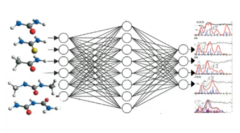
Machine learning prediction of spectra with molecular graphs
Hendrik Junkawitsch, Sandeep Kumar
Machine learning (ML) gains more importance in chemistry and may become a replacement to other computational methods such as density-functional theory. In this exercise, the participants will be exposed to some basic concepts of predicting chemical properties (e. g. X-ray spectra) with ML. The exercise will involve a gentle introduction to the types of tasks in machine learning, and a general idea of how ML algorithms work. We will then define the problem of predicting spectra with ML in detail. This will then be followed by a brief discussion and basic introduction to molecular representations such as SMILES, Coulomb matrices and graphs using python. The session will then have a discussion of message-passing on graph neural networks. The hands-on task will be training a small GNN framework that predicts spectra or other molecular properties.
Training 11
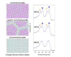
Charge Density Waves in a TaS2 surface investigated by photoelectron spectroscopy
Nomi Sorgenfrei, Danilo Kühn, Johannes Lütgert
Photoelectron spectroscopy (PES) is a powerful tool to investigate the chemical surrounding of different atoms. Not only is PES element-specific but also sensitive to variations of the electronic surrounding of the different species due to the crystal field. This technique is called Electron Spectroscopy for Chemical Analysis or ESCA.
TaS2 is a particular interesting material for this class of experiments, since it exhibits a charge density wave (CDW) which modulates the electronic surroundings of the Tantalum atoms and is reflected in the photoelectron spectrum. During the photon school, you will prepare a TaS2 sample for measurement, characterize it and investigate the phase transitions induced by cooling the sample with liquid nitrogen. You will also analyse the data, learn how to use fitting routines and prepare a short presentation.
Training 12
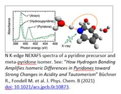
Photochemistry in Liquids – Time-resolved and steady-state soft X-ray absorption
Mattis Fondell & Leo Cordsmeier
The AXSYS-nm Transmission Near-edge X-ray absorption fine structure (NEXAFS) station is dedicated to time-resolved and steady-state soft x-ray absorption spectroscopy on liquid samples that are continuously refreshed in a flat jet system. The experiment provides the unique capability to probe electronic structure changes of molecules in solution through soft X-ray absorption on picosecond timescales subsequently to a laser excitation. This training will introduce liquid-jets. The participants will record X-ray absorption spectra of molecule in solution at the nitrogen K- and the iron L-edge. They will use a laser system to promote the molecule to a valence excited state and detect the characteristic X-ray absorption changes related to this excitation.