Department Spin and Topology in Quantum Materials
3D microscopy using the standing wave technique
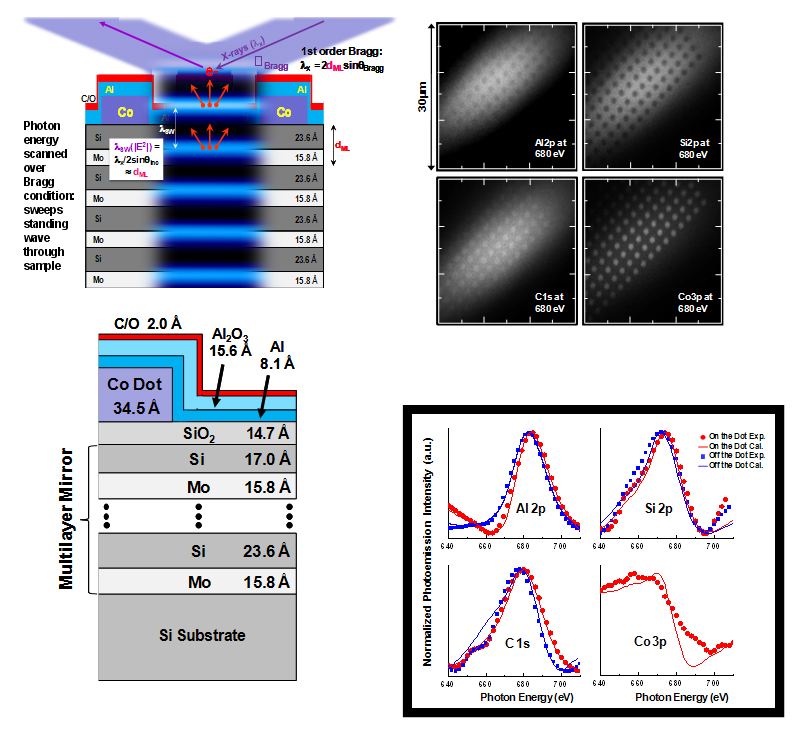
Depth resolution along the surface normal down to a few angstroms is achieved by using standing-wave excitation. In this example the sample is a Ag/Co/Au trilayer, grown on a Si/MoSi2 multilayer mirror, with the bottom Ag layer in a wedge profile. Tuning the incident x-ray to the mirror Bragg angle sets up a standing x-ray wave field in the multilayer and the trilayer wedge structure.
F. Kronast, R. Ovsyannikov, A. Kaiser, C. Wiemann, S.-H. Yang, D. E. Bürgler, R. Schreiber, F. Salmassi, P. Fischer, H. A. Dürr, C. M. Schneider, W. Eberhardt, and C. S. Fadley
Appl. Phys. Lett.: 93, 243116 (2008); 97, 062503 (2010)