Institute Electrochemical Energy Storage
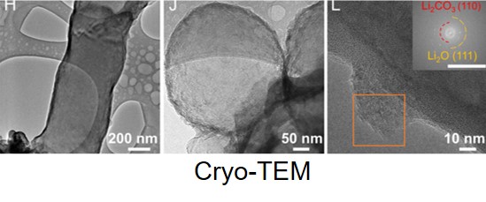
Cryo-TEM
1. Cryo-TEM and tomography
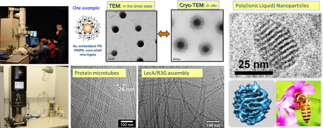
Cryogenic transmission electron microscopy (Cryo-TEM) is a method of choice to investigate the morphology of colloidal systems in situ. In our research cryo-TEM is demonstrated to be an powerful method to characterize the swollen state of microgels in solution, which is impossible to be observed by normal TEM with dried samples. In addition, the morphological evolution of polymer particle shape and interior nanostructure can be well presented with cryo-TEM and tomography.
1. W. Zhang, Z. Kochovski, Y. Lu, B. V. K. J. Schmidt, M. Antonietti, J. Yuan, ACS Nano 2016, 10, 7731-7737.
2. G. Yang, X. Zhang, Z. Kochovski, Y. Zhang, B. Dai, F. Sakai, L. Jiang, Y. Lu, M. Ballauff, X. Li, C. Liu, G. Chen, M. Jiang, J. Am. Chem. Soc. 2016, 138, 1932–1937.
3. G. Yang, H. Ding, Z. Kochovski, R. Hu, Y. Lu, Y. Ma, G. Chen, M. Jiang, Angew. Chem. Int. Ed. 2017, 56, 10691-10695.
4. R. Liu, Z. Kochovski, L. Li, Y. Yin, J. Yang, G. Yang, G. Tao, A. Xu, E. Zhang, H. M. Ding, Y. Lu, G. Chen, M. Jiang, Angew. Chem. Int. Ed. 2020, 59, 9617-9623
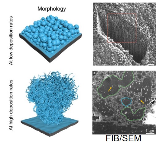
Characterization of sensitive battery/energy materials by Cryo-TEM
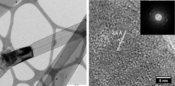
Cryogenic electron microscopy (cryo-EM) has recently emerged as a revolutionary technique for analysis of reactive and sensitive energy materials such as metal-organic frameworks (MOFs), hybrid organic-inorganic halide perovskite solar cells and battery materials at the atomic scale. These materials have previously resisted atomic-resolution studies by traditional transmission electron microscopy (TEM) because of their instability under the electron beam. Cryo-EM offers a unique way to preserve the native state of these materials by shock-freezing them under cryogenic conditions (~90 K) and to image them at nano/atomic scale.
[1] K. Dong, Y. Xu, J. Tan, M. Osenberg, F. Sun*, Z. Kochovski, D. Pham, S. Mei, A. Hilger, E. Ryan, Y. Lu*, J. Banhart, I. Manke*, ACS Energy Lett. 2021, 6, 1719–1728.

